Ultrasound in Puerto Vallarta
Everything you should know about your Ultrasound in Puerto Vallarta
What is an Ultrasound?
An ultrasound, also called sonography, is an imaging study that uses sound waves to create pictures of different body parts, organs, and tissues. In addition, with these high-frequency sound waves, an ultrasound is able to show details and motion inside our body (blood flow, heartbeats, etc.). Thanks to these detailed images, this type of imaging study can provide valuable information that can help to diagnose and treat many health conditions.
Ultrasound definition: An ultrasound is an imaging study that uses sound waves that are of such high frequency that humans are actually incapable of hearing them. Ultrasounds operate at frequencies that are above our hearing threshold, which is at approximately 20,000 hertz.
Things to note about an ultrasound scan
- Bats use ultrasound waves to visualize their preys and obstacles while in flight.
- Ultrasound is used in many different fields and applications. It is used in industrial applications, in nondestructive testing, and in the medical field.
- In medicine, ultrasound scans are widely used, safe, and affordable.
- Radiologists and doctors use ultrasound scans to diagnose and treat many different illnesses.
- Fortunately, ultrasounds are convenient imaging studies that usually do not require any special preparation beforehand.
- An important safety feature of ultrasounds is that they do not use nor produce radiation. Therefore, there is no radiation exposure and no risk of developing cancer due to radiation.
How does an ultrasound work? How does an ultrasound capture images?
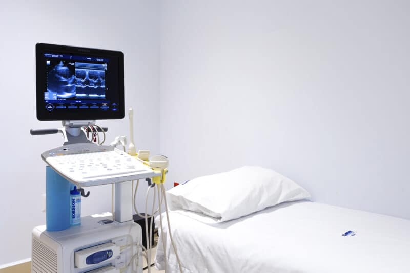
In medicine, an ultrasound, also known as medical ultrasound, is an equipment that works when a radiologist places its transducer on the patient’s skin. A transducer is a small probe through which high-frequency sound waves are sent from the ultrasound equipment into the body. Once these sound waves hit the organs and body parts, they bounce back and are collected by the transducer, which in turn sends them to the ultrasound equipment.
Finally, a computer in the ultrasound equipment uses these sound waves to create images. Accordingly, radiologists use these images to evaluate and interpret their meaning. Once this is done, radiologists give their clinical opinion regarding a possible diagnosis. Due to their usefulness and convenience, there are several types of ultrasounds that are widely used by doctors worldwide.

Types of ultrasounds
In healthcare, there are several types of ultrasounds. Therefore, ultrasounds can be classified depending on how they are performed, or according to the type of study and its end results.
External ultrasounds
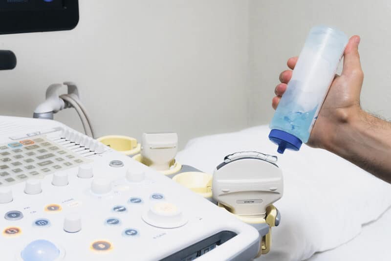
External ultrasounds are studies in which the radiologist puts lubricating gel on the patient’s skin, and then places the transducer on the gel that was recently applied. Subsequently, the radiologist will move the transducer over the parts of the body that need to be examined. Fortunately, in an external ultrasound, the patient will not feel any discomfort, as it will only feel the transducer´s movement over the skin. Common examples of external ultrasounds are pregnancy ultrasounds, liver ultrasounds, and abdomen ultrasounds.
Internal ultrasounds
This type of ultrasound is usually used to evaluate a patient’s urinary system or its internal reproductive organs. Internal ultrasounds are performed by placing the transducer inside a man´s rectum or a women´s vagina. Most often, pain medications are given to the patient prior to the study in order to reduce discomfort. Unfortunately, internal ultrasounds are not as safe or as comfortable as external ultrasounds, as they are performed inside the body. In addition, there is always a slight risk of bleeding during this type of diagnostic study. Common examples of internal ultrasounds are transvaginal ultrasounds and transrectal ultrasounds.
Doppler Ultrasounds, 2D Ultrasounds, 3D Ultrasounds, and 4D Ultrasounds
Another way of classifying ultrasounds is according to the type of ultrasound study and the images it produces. Generally speaking, there are four types of ultrasounds:
- Doppler Ultrasounds: A Doppler ultrasound is a special type of diagnostic study that uses sound waves in a different frequency to show and measure how liquids move inside the body. Most often, Doppler ultrasounds are used to measure blood flow in arteries and veins. Specifically, this type of imaging study can measure the direction and speed of blood flow.
- 2D Ultrasounds: 2d ultrasounds create cross-sectional images of organs and body parts in two-dimensional planes. In general, 2d is the standard and most commonly used ultrasound study, as it’s widely available and well known by doctors and Diagnostic Medical Sonographers.
- 3D Ultrasounds: Three-dimensional ultrasounds are made by putting several 2d images that were taken from different angles and creating a single 3d image. 3d ultrasounds are normally used for pregnancy ultrasound studies, to show a “picture” of a baby.
- 4D Ultrasounds: 4d ultrasounds create a kind of movie with a live video effect. 4d ultrasounds are the most popular kind of imaging study during pregnancy, as it provides a real-time movie of a baby inside its mother’s womb.
Are Ultrasounds Safe? Ultrasound Risk and Benefits
Since its introduction to the medical field in 1956 by Ian Donald, ultrasound scans have always had an excellent safety record. Accordingly, an ultrasound’s safety is based on its use of high-frequency sound waves which are classified as non-ionizing radiation. As opposed to X-Rays and CT Scans, which generate and use ionizing radiation (which can cause cancer), ultrasounds do not emit nor use this type of radiation. Therefore, ultrasound imaging is considered very safe when used prudently by a professional radiologist or sonographer. However, in some cases, it may cause some biological effects on the human body. Here are the main Risks and Benefits of an ultrasound:
Ultrasound risks
- Although it’s unnoticeable, ultrasound waves can heat tissue in a minimal way.
- In addition, ultrasound waves can produce cavitation, which is the effect of producing small pockets of gas in tissues and body fluids.
- The long term effects of these two factors are still unknown. Nevertheless, the FDA and the American Institute of Ultrasound in Medicine recommend ultrasound imaging to be used prudently and only when necessary.
- Due to these facts, the use of 3D and 4D ultrasound imaging for the sole purpose of obtaining pictures or videos of babies during pregnancy is not recommended.
Ultrasound benefits:
- Compared to other diagnostic studies, ultrasound is considered safe, as it produces no side effects when prudently used.
- Ultrasound scans do not expose patients to ionizing radiation, which is the kind of radiation that can cause cancer.
- Most of the studies performed using ultrasound are non-invasive, and therefore are painless and safe.
- Ultrasound scans are inexpensive when compared to other imaging studies such as CT Scans and MRIs.
- When an invasive procedure is needed, ultrasound guidance allows the doctor to have a real-time visual aid, which can make the procedure faster and safer.
- Furthermore, an ultrasound is a piece of smaller and lighter equipment that can be moved to different areas of a hospital when needed.
Ultrasound diagnostic studies have been used for over 30 years, having an excellent safety record.
How can I find an ultrasound near me in Puerto Vallarta? How can I find an urgent care with ultrasound in Puerto Vallarta?
If you need to have an ultrasound scan in Puerto Vallarta, there are several medical facilities that offer this imaging study. In this case, there are two types of medical facilities in which you could get an ultrasound scan; in private hospitals or in stand-alone imaging centers. However, if you are looking for the nearest and most convenient location to get your ultrasound scan, your best options are our three hospitals and urgent care centers which are strategically located across Banderas Bay:
Our Locations
CT Scan Studies, Urgent care center open 24/7, and Walk-in clinic.
CT Scan and Open MRI Studies, ICU, Urgent care center open 24/7, and Walk-in clinic.
CT Scan Studies, 24/7 emergency medicine, On-site specialists, and Walk-in clinic.
Ultrasound in Puerto Vallarta at Hospital CMQ City Center:
If you are staying in the southern part of Puerto Vallarta, in places such as The Malecon, Gringo Gulch, Amapas, The Romantic Zone, Conchas Chinas or Mismaloya, Hospital CMQ City Center is your nearest urgent care and imaging center. Hospital CMQ City Center´s Diagnostic Ultrasound Service is available 24 hours per day, 7 days per week.
Ultrasound in Puerto Vallarta at Hospital CMQ Premiere:
Alternatively, if you are staying in Puerto Vallarta´s central zone, in places such as Fluvial Vallarta, the Hotel Zone, Aralias, Versalles, or Marina Vallarta, Hospital CMQ Premiere will be the closest urgent care and imaging center from your location. Hospital CMQ Premiere´s Diagnostic Ultrasound Service is also available 24 hours per day, 7 days per week.
Ultrasound in Puerto Vallarta at Hospital CMQ Riviera Nayarit:
On the other hand, if you are living or staying in Puerto Vallarta´s beautiful northern shores, in places such as Bucerias, La Cruz de Huanacaxtle, Sayulita, or Punta Mita, Hospital CMQ Riviera Nayarit is definitely your nearest urgent care and ultrasound alternative. For your convenience, our urgent care and Diagnostic Ultrasound Service is located in Bucerias, Nayarit, and is available 24 hours per day, 7 days per week.
How can I say Ultrasound in Spanish? If you are in Puerto Vallarta and need this type of study, it will be useful to know the Spanish word for an Ultrasound scan. The Spanish word for CT scan is “Ultrasonido” or “Estudio de Ultrasonido”.
How much does an Ultrasound cost in Puerto Vallarta?
In Mexico, and in Puerto Vallarta in particular, ultrasound prices can vary depending on the hospital and/or imaging center where the study is performed. Moreover, ultrasound prices will also fluctuate depending on whether it’s a scheduled or an emergency procedure if it’s a holiday or a weekend. Nevertheless, for your information and convenience, we have included in the table below the average price range of the most common ultrasound scans at Hospital CMQ in Puerto Vallarta:
| Ultrasound Scan | Ultrasound Price Range | (Approximate cost range for scheduled outpatient studies. Studies performed on an emergency basis have a higher cost). |
|---|---|---|
| Liver ultrasound / Gallstones ultrasound | $995.00 Mexican pesos | $1,795.00 Mexican pesos |
| Thyroid ultrasound | $995.00 Mexican pesos | $1,795.00 Mexican pesos |
| Breast ultrasound | $995.00 Mexican pesos | $1,795.00 Mexican pesos |
| Gallbladder ultrasound | $995.00 Mexican pesos | $1,795.00 Mexican pesos |
| Renal ultrasound / Kidney ultrasound | $995.00 Mexican pesos | $1,795.00 Mexican pesos |
| Ectopic pregnancy ultrasound | $995.00 Mexican pesos | $1,795.00 Mexican pesos |
| Testicular ultrasound | $995.00 Mexican pesos | $1,795.00 Mexican pesos |
| Prostate ultrasound | $995.00 Mexican pesos | $1,795.00 Mexican pesos |
| Pregnancy ultrasound | $1,137.00 Mexican pesos | $1,937.00 Mexican pesos |
| Gender ultrasound | $1,137.00 Mexican pesos | $1,937.00 Mexican pesos |
| Abdominal ultrasound | $1,279.00 Mexican pesos | $2,079.00 Mexican pesos |
| Transvaginal ultrasound | $1,421.00 Mexican pesos | $2,421.00 Mexican pesos |
| Pelvic ultrasound | $1,421.00 Mexican pesos | $2,421.00 Mexican pesos |
| Vaginal ultrasound | $1,421.00 Mexican pesos | $2,421.00 Mexican pesos |
| Endometriosis ultrasound | $1,421.00 Mexican pesos | $2,421.00 Mexican pesos |
| Ovarian cancer ultrasound | $1,421.00 Mexican pesos | $2,421.00 Mexican pesos |
| Uterus ultrasound | $1,421.00 Mexican pesos | $2,421.00 Mexican pesos |
| Abdominal Doppler ultrasound | $3,267.00 Mexican pesos | $4,267.00 Mexican pesos |
| Leg doppler ultrasound scan (one leg) | $3,551.00 Mexican pesos | $4,551.00 Mexican pesos |
| Arterial Doppler ultrasound | $3,551.00 Mexican pesos | $4,551.00 Mexican pesos |
| Carotid ultrasound | $3,835.00 Mexican pesos | $4,835.00 Mexican pesos |
| Carotid doppler ultrasound | $3,835.00 Mexican pesos | $4,835.00 Mexican pesos |
| Heart ultrasound | $7,540.00 Mexican pesos | $10,540.00 Mexican pesos |
Special pricing available for hotels, restaurants and corporations.
How should I prepare for an Ultrasound in Puerto Vallarta?
If you need to have an ultrasound scan, it will probably be because your doctor ordered it. Fortunately, at Hospital CMQ we have a professional team of radiologists, sonographers, and nurses who specialize in ultrasound diagnostic studies.
First of all, you should know: How is an ultrasound performed?
An external ultrasound is performed in the following way: Our radiologist or one of our sonographers will apply a special lubricating gel on your skin. This lubricating gel helps to transmit the ultrasound´s sound waves and also reduces friction. Subsequently, the transducer will send high-frequency waves through the part of your body in which the study is being performed. In turn, these sound waves will bounce back (they will produce an echo) when they hit a dense or solid object, like a bone, a blood vessel, or an organ. Accordingly, these echoes are captured by the transducer and are transmitted to the ultrasound´s computer. Finally, once the study has been performed, our radiologist will clean the gel from your skin, interpret the final images, and will write his clinical opinion in a medical report. This medical report and the captured images will be given both to you and your doctor.
If you would like to know how an internal ultrasound is performed, we recommend that you talk to your doctor about it, as this is a procedure that is more complex and that involves certain minor risks.
Things to know before, during, and after my ultrasound in Puerto Vallarta
Things to know before your ultrasound scan
For most ultrasound scans, especially for external ultrasounds performed in the abdominal region, you will need to drink four glasses of water (32 ounces), one hour before your exam. If during this time, you feel the need to go to the bathroom, you may do so, but you should keep drinking water to replenish this liquid. In addition, for this type of ultrasounds, you will need to refrain from eating or drinking any food or substance for eight hours prior to your exam. Furthermore, if you have any jewelry or valuables, we would ask that you leave them home or deposit them in the hospital´s safe box.
Finally, please note that we would like to make your exam and waiting time as pleasant as possible. Therefore, please consider bringing comfortable clothing, and your favorite book or magazine. Last but not least, something that is very important: if your doctor gave you an ultrasound order, please bring it with you.
What will happen during your ultrasound scan?
Once you arrive at our imaging facility, our front desk staff will ask you for your name, your ultrasound order, and/or the type of ultrasound that you require. Next, our radiologist or one of our sonographers will ask you to change into a hospital gown. Subsequently, you will be taken to the ultrasound room where your study will be performed, and our radiologist will answer any questions that you may have. Once you are ready and there are no further doubts, you will be asked to lay down on the exam table and our radiologist will apply the lubricating gel on the region to be studied. After this, our radiologist will place the ultrasound´s transducer on your skin with the lubricating gel, and the transducer will transmit and collect high-frequency sound waves. Simultaneously, as the sound waves travel through your body, they will bounce back when they hit dense organs or tissue in your body. Finally, the transducer will capture these images and will send them to the ultrasound´s computer.

Your Ultrasound’s interpretation and report
By the end of your study, the moving and detailed images will be recorded and analyzed by our radiologist. In addition, he will write your medical report based on the captured images and his clinical interpretation. Accordingly, our radiologist will give you and your doctor the corresponding images and medical reports. Most importantly, your ultrasound study, its images, and results will be available online through our digital PACS system. This way, you or the doctors that you authorize will have access to your study from anywhere, at any time. In total, you can expect your ultrasound scan to take anywhere from 35 to 50 minutes.
Our radiologist, Dr. Cesar Medina, will give you a CD containing your ultrasound images, and his clinical interpretation which will be included in his medical report. In addition, he will be available to answer any questions that you may have.
What should I do after my ultrasound?
As previously mentioned, after your exam, our radiologist will give you your medical report and a CD with your ultrasound images. In addition, he will send a copy of these items to your doctor or treating physician. Furthermore, if you have any questions related to your ultrasound exam, please call us at (322) 22 66500. Similarly, you can call us at this number to request a copy of your images and your medical report. Fortunately, other than this, there is normally nothing that you should do after your sonography exam.
Ultrasound pictures
Pregnancy ultrasound images/baby ultrasound pictures

5 week ultrasound
Through this 5-week pregnancy ultrasound, we can observe a gestational sac containing an embryo. Similarly, the embryo´s cardiac frequency can also be obtained and recorded. In this case, the embryo’s normal cardiac frequency would be between 100 and 180 beats per minute. In addition, the mother will be able to listen to its baby’s heartbeat even at this early stage.

13 week ultrasound
During a 13-week pregnancy ultrasound, the radiologist is able to measure the size of the fetus. Among healthcare providers, this measure is known as the “Crown-rump length (CRL)”, which is the length of a human fetus from the top of its head to its buttocks. For example, the fetus in this image has a CRL measurement of 6.7 centimeters. In addition, the radiologist will check the fetus’s legs, its cardiac activity, and any signs of alterations such as Down syndrome.

20 week ultrasound
On the image on the left, we can observe the baby’s abdominal development. In contrast, in the image on the right we can observe a mature placenta and the amount of liquid surrounding it. Likewise, the renal arteries can be observed and examined in order to determine the health and development of the baby’s kidneys.
Breast cancer ultrasound images
Examples of breast ultrasounds used to detect breast cancer

Breast ultrasound suggesting a probable cyst or tumor:
In this 2d (two-dimensional) breast ultrasound we can observe images of the right breast of a young female. In this case, the black color that is present in the patient’s breast indicates the probable existence of an oval-shaped tumor or a cyst. Due to the size of the oval shape and the presence of several color tones, the suspicion of breast cancer becomes a probable possibility. Therefore, a breast biopsy will be necessary in order to confirm or reject the existence of breast cancer.

Breast ultrasound showing the probable existence of a malign breast cancer tumor:
This breast ultrasound shows the probable existence of a malign breast cancer lesion. In this study, the lesion’s borders are not well defined which means that the ultrasound’s sound waves are not able to travel through this area. Similarly, we can observe that the mammary tissue has been affected and “disordered” by the tumor. As in the previous example, a breast biopsy will be needed to confirm the presence of the tumor, and determine its stage of development.

Ultrasound-guided breast biopsy
Thanks to the accuracy of ultrasound imaging, it is possible to perform ultrasound-guided breast biopsies on suspicious lesions to confirm or reject the presence of illnesses such as breast cancer. In this image, we can observe a needle and its trajectory as it is inserted through the breast. This biopsy needle is used to extract and collect a small breast tissue sample. In turn, this breast tissue sample is examined to determine the existence or inexistence of breast cancer.
Ultrasound frequently asked questions
Although some medical providers might suggest getting your first ultrasound scan even earlier, our recommendation is to get your first pregnancy ultrasound at 12 weeks. This is due to the fact that the basic anatomy of your baby is visible at 12 weeks. In addition, this is the perfect time for determining your due date, checking basic anatomy, screening for Down syndrome, and other rare conditions.
Although it is recommended that your ultrasound images are evaluated and interpreted by your radiologist or gynecologist, in general terms we can say the following: While ultrasound images of a normal breast have a uniform grey color within the breast area, images of a tumor in the breast look like a black dot in the shape of a black star. However, it is important to note that even though an image of a tumor might be present in a study, it may not be a cancerous tumor. Therefore, further tests would be needed to determine if this image resembling a black star is a benign tumor or a malignant tumor (cancerous tumor).
The duration of an ultrasound scan can vary depending on the type of ultrasound needed. However, most ultrasound studies will take from 35 minutes to 50 minutes.
We would recommend getting a minimum of three ultrasounds during your pregnancy, one every three months. Specifically, we would recommend getting your three pregnancy ultrasounds in the following week:
- First pregnancy ultrasound, at week 12.
- Second pregnancy ultrasound at week 22.
- Thirds pregnancy ultrasound at week 35.
Fortunately, you will be able to see the embryo of your baby starting at week 5 of your pregnancy. At his stage, you will be able to see the basic framework and foundation of your baby, and it will be when the important systems of its body are formed. On the other hand, you will be able to see your baby itself, with beautiful images of its face and body from week 28 and onwards.
You can begin to hear your baby’s heartbeat from week 5 of your pregnancy.
A transvaginal ultrasound, also known as an endovaginal ultrasound, is one of several types of pelvic ultrasounds. A transvaginal ultrasound is by definition an internal examination. Thus, the term transvaginal literally means “through the vagina”. In a transvaginal ultrasound, the transducer (small probe) is placed 2 to 3 inches inside the vaginal canal. In general, doctors use this study to examine the female reproductive organs.
Although an ultrasound can detect tumors that may be cancerous, it will not provide a definitive answer on whether a lump or mass is cancerous or not. In this sense, it is worth noting that ultrasound images are not as exact or as detailed as MRI images or CT scan images. In addition, an ultrasound scan can have some limitations as its waves cannot go through the air (air in your lungs) nor through bones.
Fortunately, you can eat before most ultrasound studies. The only exception would be if you are having an abdomen ultrasound. In the case of an abdomen ultrasound, you cannot eat before this exam.
Your doctor might ask you to have an ultrasound after a CT scan, in some cases in which the images produced by the CT scan are not able to reflect the illness or condition that your doctor is suspecting. For example, an ultrasound may be needed after a CT scan in the case where a physician is suspecting possible kidney stones, or if a doctor needs to see the blood flow in an artery or vein.